ANATOMOFISIOLOGIA DA MAO
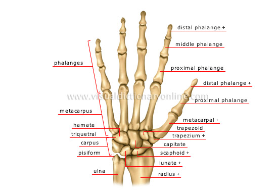
Ossos
Os ossos da mão são os ossos do carpo, os metacarpos
e as falanges, dividindo-se estas ultimas em proximais, intermédias e distais.Os
metacarpos e falanges numeram-se de 1 a 5 a partir do polegar


Ossos do carpo

Escafoide
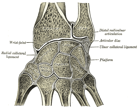
Articulações da mão
Textos
gerais
Articulação
carpo-metacarpica

Há cinco articulaçoes carpometacarpicas em cada mao

Ligações
intercarpicas

Ligamento
transverso do metacarpo

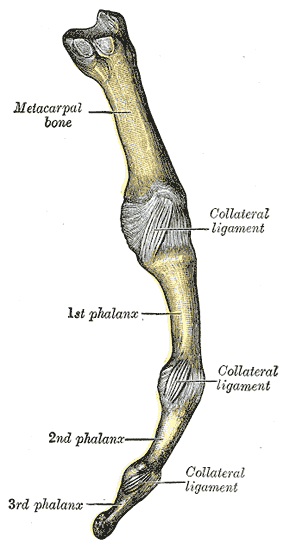
Ligações interfalangicas

Músculos da mão
Músculos extrínsecos
Os músculos extrínsecos da mão são os
que têm origem no antebraço e os tendões se inserem na mão
Uma
banda forte de tecido conjuntivo fibroso (ligamento
anular anterior do carpo) cobre os tendões e mantem-nos no seu lugar à volta
do punho
Músculos intrínsecos
Têm
origens e inserções na mão
Duas
eminências, tenar e hipotenar permitem dividir os músculos
em tenares e hipotenares
A
eminência tenar é uma saliência arredondada na base do punho e a hipotenar na
base do mínimo
Temos
de considerar ainda os músculos da palma da mão
Compreendem
os principais abdutores, adutores e opositores dos dedos
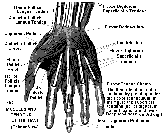
When the skin, palmar aponeurosis and flexor
retinaculum are removed, the tendons of the flexor digitorum superficialis can
be seen. Medial to the tendons is a group of muscles that act on the little
finger, the hypothenar muscles. Lateral to the tendons is a group of muscles
that act on the thumb (pollux), the thenar muscles. These two muscle groups are
covered with deep fascia.
The intrinsic muscles of the hand can be arranged into
three groups according to either to a region or to depth. Regional groups of muscles are the thenar and hypothenar group. The thenar muscles are three in number and act on the thumb. The hypothenar group are three in number and act on the little finger.
Sem comentários:
Enviar um comentário