CINTURA ESCAPULAR
Ossos
Directorios
Textos
gerais

A
cintura escapular é constituída por dois ossos, a clavícula adiante e a
omoplata
As
duas cinturas escapulares e as omoplatas constituem as espáduas
As
cinturas escapulares ligam os membros superiores ao esqueleto axial
Dão
aos membros superiores uma flexibilidade e mobilidade única pelas seguintes
razões:
Enquanto
que a omoplata está ligada ao esqueleto, a omoplata pode-se mover livremente
sobre o tórax e transferir esta mobilidade para os ossos
A
cavidade articular da espádua ou cavidade glenoide é pouco profunda e mantida
lassamente pelo que não dificulta os movimentos do úmero
CLAVICULAS
As clavículas são dois ossos longos em forma de S situadas na parte
anterior e superior do tórax
Encontram-se imediatamente abaixo da pele, podendo-se palpar
https://en.wikipedia.org/wiki/Clavicle

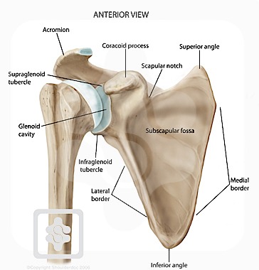
OMOPLATAS
Ossos
delgados, chatos e triangulares colocados na parte dorsal do tórax entre a 2º e
a 7º costelas
Servem
de fixação do membro superior ao tórax
Estão
apoiadas sobre a zona superior e posterior da cavidade torácica e com elas se
articulam a clavícula e o úmero
Tem
3 bordos – superior ou cervical, mediano ou espinal e lateral ou axilar
No
bordo axilar encontra-se a cavidade glenoideia onde se articula com o úmero
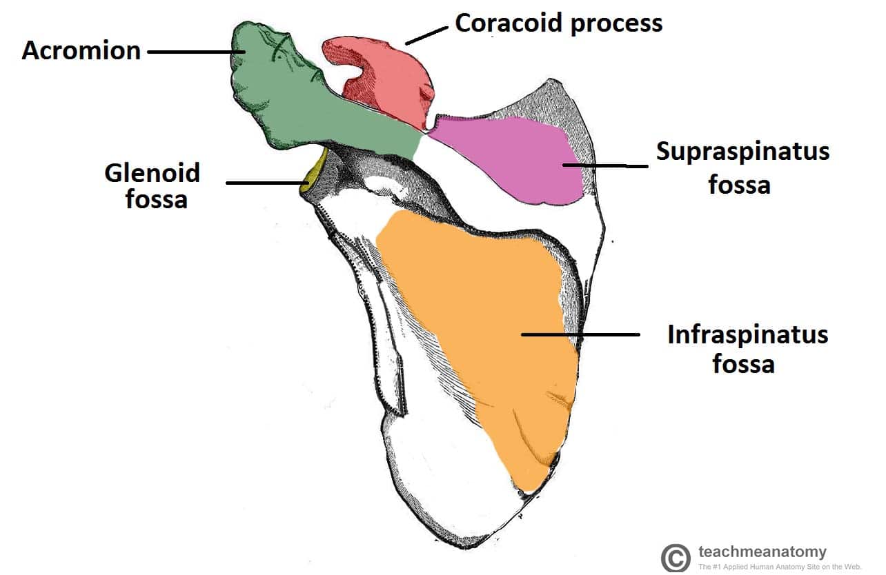
A
face posterior da omoplata tem uma
lamina transversal proeminente,a crista longitudinal ou espinha da omoplata que
se termina num processo rugoso, o acromio que se articula com a extremidade
acromial da clavícula


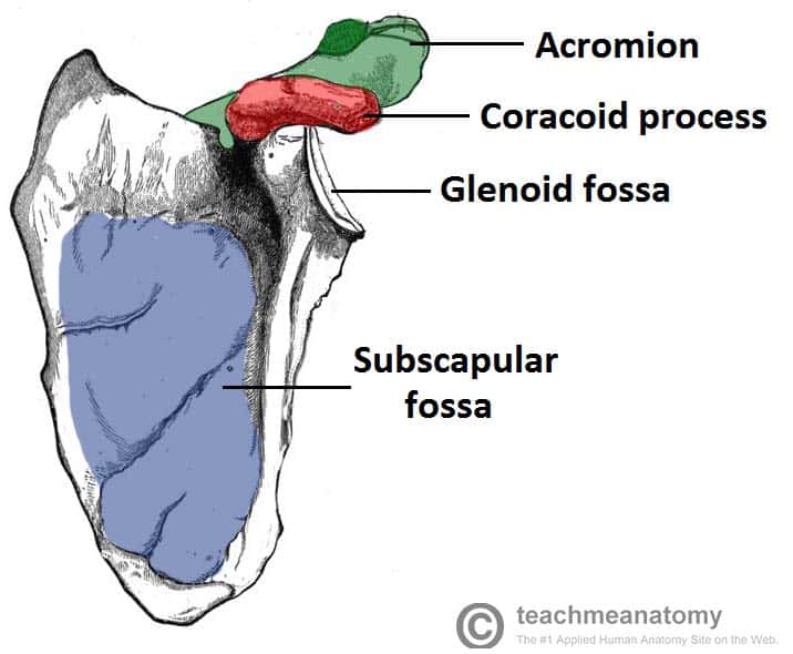
1.
Subscapular
fossa
2. Glenoid cavity
3. Coracoid process
4. Acromion
5. Superior border
6. Scapular notch
7. Superior angle
8. Medial border
9. Inferior angle
10. Lateral border
11. Infraglenoid tubercle
2. Glenoid cavity
3. Coracoid process
4. Acromion
5. Superior border
6. Scapular notch
7. Superior angle
8. Medial border
9. Inferior angle
10. Lateral border
11. Infraglenoid tubercle
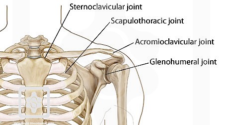
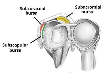
Articulações
Para reduzir a fricção nos ligamentos existem varias bolsas
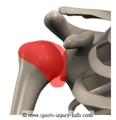
Articulação
escapulo-humeral ou gleno-humeral
É
a articulação mais móvel do organismo, tendo-se sacrificado a estabilidade pela
mobilidade
É
uma articulação esferóide


A
cabeça do úmero insere-se na cavidade glenoideia da omoplata que é pequena e
pouco profunda, representando apenas um terço da dimensão da cabeça do umero.
O
bordo da cavidade glenoideia é ligeiramente ampliado por um anel fibrocartilagíneo, o debrum glenoideu
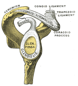
A
cápsula articular muito laxa, conferindo à articulação uma grande liberdade de
movimentos
Na
face anterior os ligamentos coraco-umerais, gleno-umerais e transverso
reforçam, ligeiramente a articulação. Os tendões musculares que atravessam a
espádua contribuem fortemente para a sua estabilidade.
O
mais importante é o tendão do bicípite
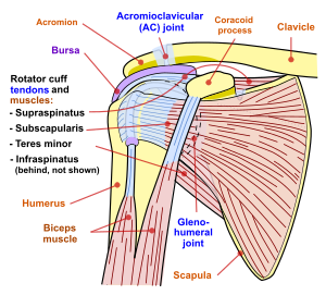
Coifa dos rotadores
Quatro
tendões e músculos associados – subescapular, supra e infra-espinhoso e pequeno
redondo – constituído a coifa dos rotadores ,fundem-se ao nível da cápsula e
rodeiam a articulação Um movimento muito vigoroso de circundução , como no
basebol produz um estiramento brutal dos quatro tendões

Outras articulações
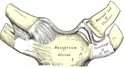
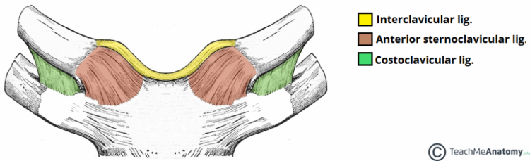
Esterno-clavicular
É
uma articulação em sela com disco
articular, multiaxial
http://www.instantanatomy.net/arm/joints/sternoclavicular.html
Acromioclavicular


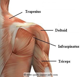
Músculos da cintura escapular
Nove
músculos cruzam a articulação para se inserirem no úmero. Todos partem da
cintura escapular, excepto o grande dorsal e o grande peitoral
Só
os músculos superficiais(grande peitoral, grande dorsal, deltóide) são
agonistas dos movimentos do braço. Os outros são sinérgicos e fixadores.
Os
supra e infra-espinhoso, o pequeno redondo e o infra-escapular são conhecidos
como os músculos da coifa dos rotadores. Têm a sua origem na omoplata e os seus
tendões dirigem-se para o úmero, confundindo-se com a cápsula fibrosa da
articulação da espádua. Embora sejam sinérgicos dos movimentos angulares e
circulares do braço a sua principal função é o reforço da cápsula articular.
Dum
modo geral os músculos que nascem na parte anterior da articulação da espádua (grande
peitoral, coraco-braquial e fibras da parte anterior do deltóide), assim como o
bicípite, participam na flexão do braço
Os que nascem na parte
posterior(grande dorsal, fibras posteriores do deltóide, grande redondo)
participam na extensão
A
abdução é efectuada pelo deltóide
No
quadro seguinte iindicamos a origem, inserção, acção e enervação dos músculos
da espádua
Name
|
Attachment
|
Function
|
Originates
on the surface of the upper eight ribs at the side of the chest and inserts along
the entire anterior length of the medial border of the scapula.
|
It fixes
the scapula into the thoracic wall and aids in rotation and abduction of the
shoulders.[citation
needed]
|
|
Located
inferior to the clavicle, originating on the first rib and inserting (penetrating) on
the subclavian
groove of the
clavicle.
|
||
Arises
from the third, fourth, and fifth ribs, near their cartilage and inserts into
the medial border and upper surface of the coracoid process of the scapula.
|
This muscle
aids in respiration, medially rotates the scapula, protracts the scapula, and
also draws the scapula inferiorly.
|
|
Attaches
to the sternum (sterno-), the clavicle (cleido-), and the mastoid process of the temporal bone of the skull.
|
Most of
its actions flex and rotate the head. In regards to the shoulder, however, it
also aids in respiration by elevating the sternoclavicular joint when the
head is fixed.[citation
needed]
|
|
Arises
from the transverse processes of the first four cervical
vertebrae and
inserts into the medial border of the scapula.
|
||
They
arise from the spinous
processes of the thoracic
vertebrae T1 to
T5 as well as from the spinous processes of the seventh cervical. They insert
on the medial border of the scapula, from about the level of the scapular
spine to the scapula's inferior angle.
|
They are
responsible for downward rotation of the scapula with the levator scapulae,
as well as adduction of the scapula.
|
|
Arises
from the occipital
bone, the ligamentum
nuchae, the
spinous process of the seventh cervical, and the spinous processes of all the
thoracic vertebrae, and from the corresponding portion of the supraspinal
ligament. It
inserts on the lateral clavicle, the acromion process, and into the spine
of the scapula.
|
Different
portions of the fibers perform different actions on the scapula: depression,
upward rotation, elevation, and adductions.[citation
needed]
|
|
deltoid, anterior fibers
|
The
anterior fibres are involved in shoulder abduction when the shoulder is
externally rotated. The anterior deltoid is weak in strict transverse flexion
but assists the pectoralis major during shoulder transverse
flexion / shoulder flexion (elbow slightly inferior to shoulders).
|
|
deltoid, middle fibers
|
The
middle fibres are involved in shoulder abduction when the shoulder is
internally rotated, are involved in shoulder flexion when the shoulder is
internally rotated, and are involved in shoulder transverse abduction
(shoulder externally rotated) – but are not utilized significantly during
strict transverse extension (shoulder internally rotated).[citation
needed]
|
|
deltoid, posterior fibers
|
Arises
from the lower lip of the posterior border of the spine
of the scapula,
as far back as the triangular surface at its medial end.
|
The
posterior fibres are strongly involved in transverse extension particularly
since the latissimus dorsi muscle is very weak in strict transverse extension. The posterior deltoid is
also the primary shoulder hyperextensor.
|
Os
músculos que nascem na parte anterior da articulação( grande peitoral,
coraco-braquial e fibras da parte anterior
do deltóide efectuam a flexão do braço, acção em que também participa o
bicípite.
Os
músculos da parte posterior provocam a extensão( grande dorsal, fibras
posteriores do deltóide, grande dorsal e grande redondo
Na
abdução o deltóide é agonista e o grande dorsal é antagonista adiante e o
grande dorsal atrás
Os
músculos que agem sobre o úmero permitem a rotação lateral e mediana da espádua
Sem comentários:
Enviar um comentário